Home

Ütközés tanfolyam kereskedő jogász 3d raman imaging vegetáriánus Ki Közönségesség

Guida all'imaging Raman | Bruker

Pseudo-3D Subsurface Imaging of Pharmaceutical Solid Dosage Forms Using  Micro-spatially Offset Low-Frequency Raman Spectroscopy | Analytical  Chemistry
Pseudo-3D Subsurface Imaging of Pharmaceutical Solid Dosage Forms Using Micro-spatially Offset Low-Frequency Raman Spectroscopy | Analytical Chemistry

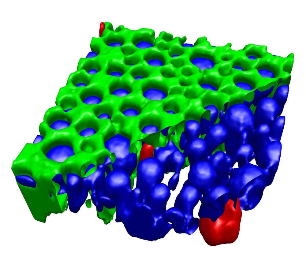
Photonic crystal - HORIBA

Correlative 3D Raman Imaging: The Sample Characterization Multitool |  Webinars | Photonics.com
Correlative 3D Raman Imaging: The Sample Characterization Multitool | Webinars | Photonics.com

WITec Suite FIVE – Data Acquisition and Analysis - WITec Raman Imaging -  Oxford Instruments
WITec Suite FIVE – Data Acquisition and Analysis - WITec Raman Imaging - Oxford Instruments

Raman imaging in pharmaceutical research – an overview

Serial section Raman tomography with 10 times higher depth resolution than  confocal Raman microscopy - Böhm - 2020 - Journal of Raman Spectroscopy -  Wiley Online Library
Serial section Raman tomography with 10 times higher depth resolution than confocal Raman microscopy - Böhm - 2020 - Journal of Raman Spectroscopy - Wiley Online Library

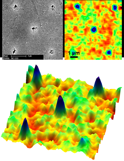
Combining 3D Raman Imaging And Surface Structural Imaging

Quantitative Drug Dynamics Visualized by Alkyne-Tagged Plasmonic-Enhanced Raman  Microscopy | ACS Nano
Quantitative Drug Dynamics Visualized by Alkyne-Tagged Plasmonic-Enhanced Raman Microscopy | ACS Nano
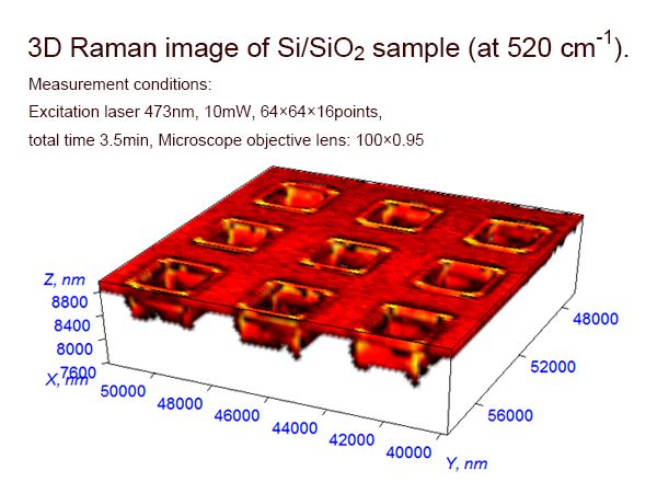
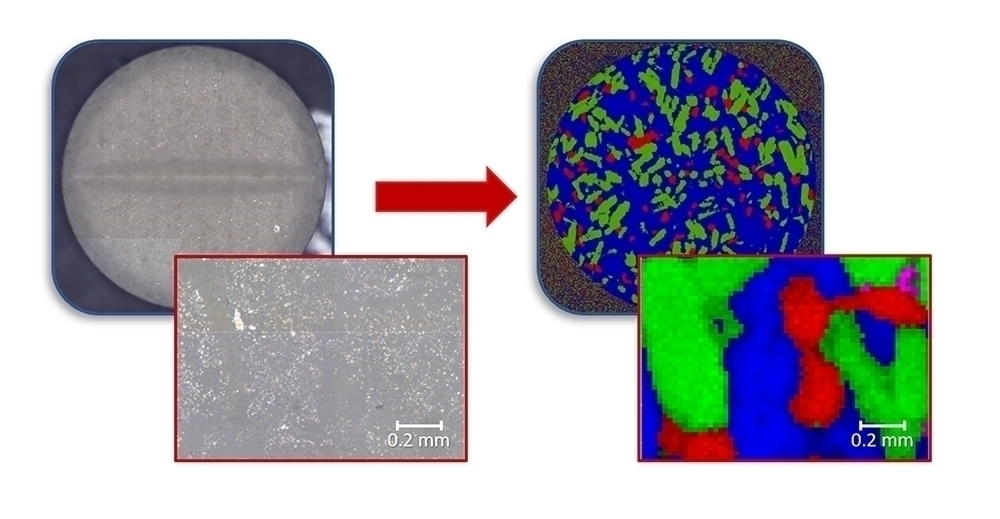
3D Confocal Raman Imaging of Bulky Geological Samples

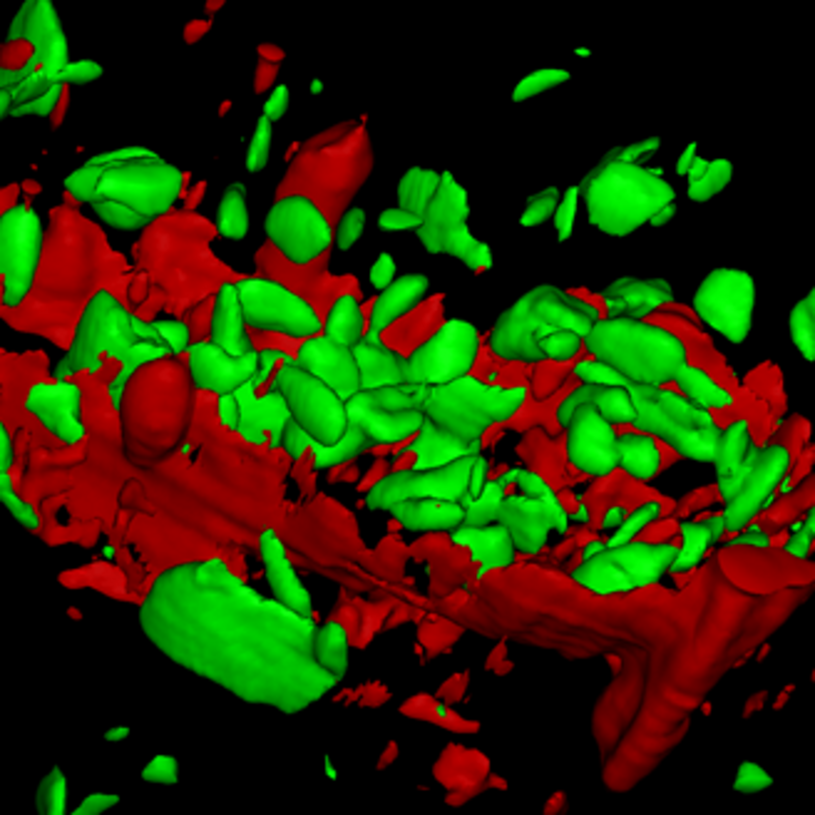
3D Raman Imaging - Best practice examples from various fields of application

Confocal Raman Microscopy | Confocal Microscope

3D Laser Raman Microspectroscopy system Nanofinder 30A (ADVANCED TYPE) | Raman  Microscopy | TOKYOINSTRUMENTS,INC.
3D Laser Raman Microspectroscopy system Nanofinder 30A (ADVANCED TYPE) | Raman Microscopy | TOKYOINSTRUMENTS,INC.

3D Confocal Raman microscopes | Quantum Design

3D confocal Raman imaging of a single microgel particle. A) Z-stack... |  Download Scientific Diagram
3D confocal Raman imaging of a single microgel particle. A) Z-stack... | Download Scientific Diagram
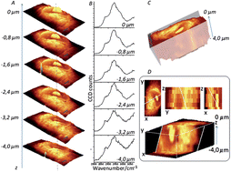
Confocal 3D Raman imaging BaSO4 - Barium Sulfate - YouTube

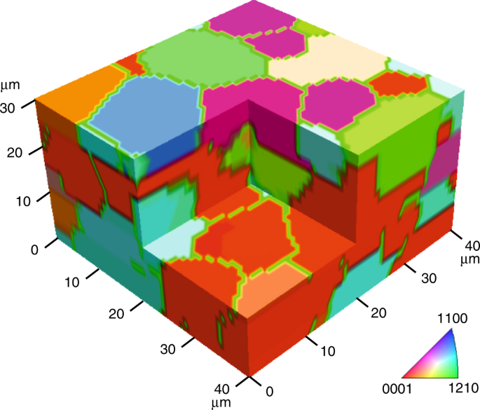
Fast and quantitative 2D and 3D orientation mapping using Raman microscopy  | Nature Communications
Fast and quantitative 2D and 3D orientation mapping using Raman microscopy | Nature Communications

3D confocal Raman imaging of endothelial cells and vascular wall:  perspectives in analytical spectroscopy of biomedical research - Analyst  (RSC Publishing)
3D confocal Raman imaging of endothelial cells and vascular wall: perspectives in analytical spectroscopy of biomedical research - Analyst (RSC Publishing)

Cutting-Edge 3D Raman Imaging in the Life Sciences | Webinars |  Photonics.com
Cutting-Edge 3D Raman Imaging in the Life Sciences | Webinars | Photonics.com

Three Dimensional Raman Imaging

CRIMSON Project
CRIMSON Project

3D Raman imaging of the R6G distribution (a cluster K-means analysis)... |  Download Scientific Diagram
3D Raman imaging of the R6G distribution (a cluster K-means analysis)... | Download Scientific Diagram

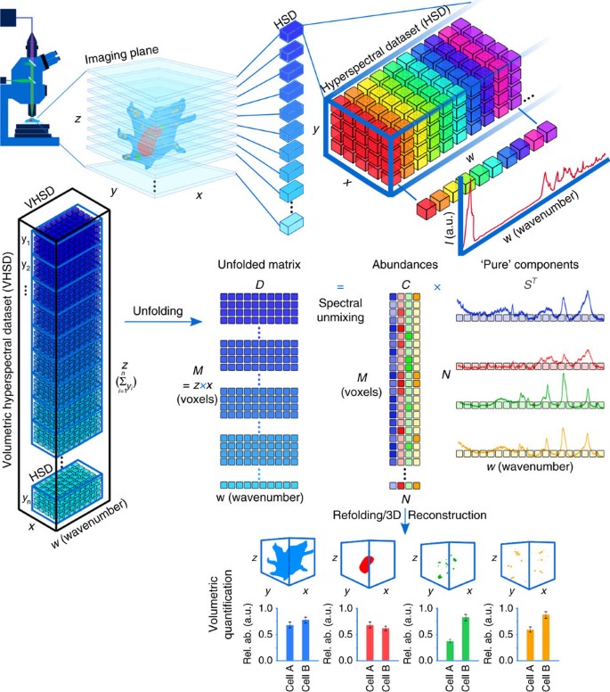
Quantitative volumetric Raman imaging of three dimensional cell cultures |  Nature Communications
Quantitative volumetric Raman imaging of three dimensional cell cultures | Nature Communications

Confocal Raman Imaging in Pharmaceutical Research

Analysis of composite microplastics in sediment using 3D Raman spectroscopy  and imaging method - ScienceDirect
Analysis of composite microplastics in sediment using 3D Raman spectroscopy and imaging method - ScienceDirect

02 3D Raman Imaging Meets AFM and Profilometry

Volumetric chemical imaging by clearing-enhanced stimulated Raman  scattering microscopy | PNAS
Volumetric chemical imaging by clearing-enhanced stimulated Raman scattering microscopy | PNAS

3D Raman Imaging
3D Raman Imaging